Home

deprem düğme çene standing knee x ray arazi bütçe pruva

Normal knee x-rays | Radiology Case | Radiopaedia.org

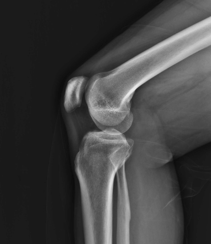
Radiographic optimization of the lateral position of the knee joint aided  by CT images and the maximum intensity projection technique | Journal of  Orthopaedic Surgery and Research | Full Text
Radiographic optimization of the lateral position of the knee joint aided by CT images and the maximum intensity projection technique | Journal of Orthopaedic Surgery and Research | Full Text

X-Knee
X-Knee

Knee standing.wmv
Knee standing.wmv

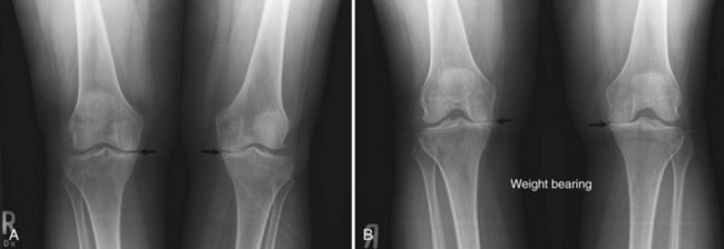
A. Long-standing X-ray showing unicompartmental medial knee... | Download  Scientific Diagram
A. Long-standing X-ray showing unicompartmental medial knee... | Download Scientific Diagram

The utility of standing knee radiographs for detection of lipohemarthrosis:  comparison with supine horizontal beam radiographs | European Radiology
The utility of standing knee radiographs for detection of lipohemarthrosis: comparison with supine horizontal beam radiographs | European Radiology

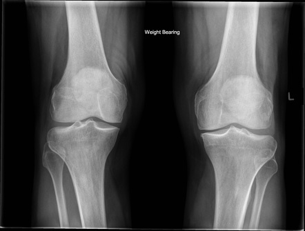
Knee (AP weight-bearing view) | Radiology Reference Article |  Radiopaedia.org
Knee (AP weight-bearing view) | Radiology Reference Article | Radiopaedia.org

The X-Ray Knee instability and Degenerative Score (X-KIDS) to determine the  preference for a partial or a total knee arthroplasty (PKA/TKA)
The X-Ray Knee instability and Degenerative Score (X-KIDS) to determine the preference for a partial or a total knee arthroplasty (PKA/TKA)

Premium Photo | X-ray knee joint (standing view) finding degeneratine  change of left knee on red mark.
Premium Photo | X-ray knee joint (standing view) finding degeneratine change of left knee on red mark.

The importance of a standing knee X-ray to evaluate arthritis – Steve  Meyers, MD
The importance of a standing knee X-ray to evaluate arthritis – Steve Meyers, MD

Knee X-Ray: Anatomy, Procedure & What to Expect

X-ray of a Columbus knee: a.p. view pre- and postoperatively (standing... |  Download Scientific Diagram
X-ray of a Columbus knee: a.p. view pre- and postoperatively (standing... | Download Scientific Diagram

Standing X-Ray Versus MRI

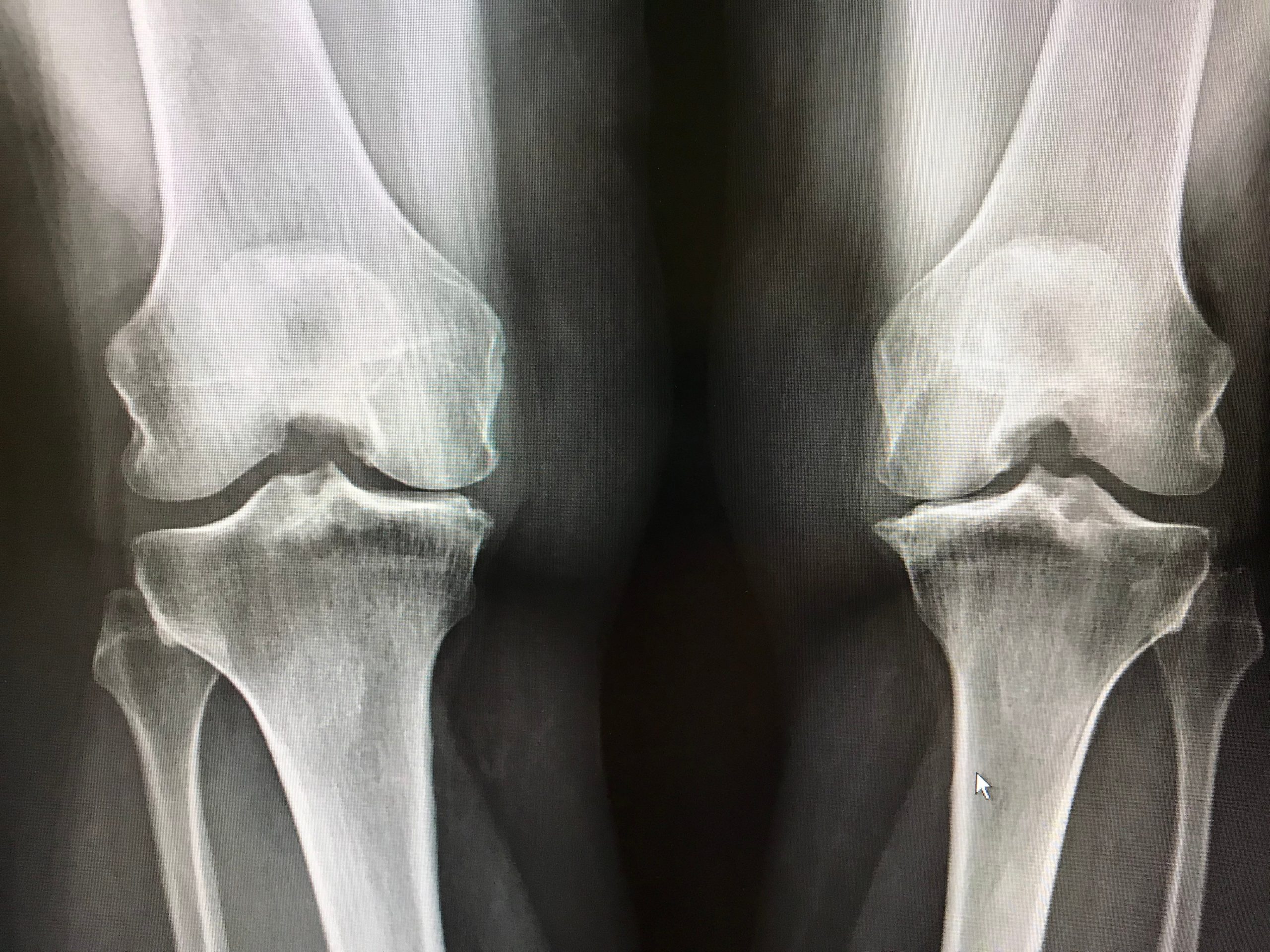
standing both knee views x-ray show normal joint Stock Photo | Adobe Stock

Radiographic Positioning Examples of the Leg and Knee - CE4RT

Anteroposterior X-ray of both knees in the standing position on patient...  | Download Scientific Diagram
Anteroposterior X-ray of both knees in the standing position on patient... | Download Scientific Diagram

Ortho Dx: "Popping" and pain in an adolescent's knee - Clinical Advisor

X-ray Right Knee AP Lateral Standing View | Test Price in Delhi | Ganesh  Diagnostic
X-ray Right Knee AP Lateral Standing View | Test Price in Delhi | Ganesh Diagnostic

x ray knee joint ap lateral view | x ray knee standing | x ray knee  positioning | AP weight bearing - YouTube
x ray knee joint ap lateral view | x ray knee standing | x ray knee positioning | AP weight bearing - YouTube

Knee Imaging Techniques and Normal Anatomy | Musculoskeletal Key

Knee X-ray의 Anatomy와 Positioning : 네이버 블로그

PDF] The one-leg standing radiograph | Semantic Scholar

Getting the perfect lateral knee on the first try is super rewarding, but  when the image isn't perfect it can be difficult to know if the knee is  rotated too much or
Getting the perfect lateral knee on the first try is super rewarding, but when the image isn't perfect it can be difficult to know if the knee is rotated too much or

Knee AP View X-ray - Aarthi Scans and Labs

X-ray P.A view both knee standing position. | Download Scientific Diagram

Difference in joint line convergence angle between the supine and standing  positions is the most important predictive factor of coronal correction  error after medial opening wedge high tibial osteotomy | Knee Surgery,
Difference in joint line convergence angle between the supine and standing positions is the most important predictive factor of coronal correction error after medial opening wedge high tibial osteotomy | Knee Surgery,

Knee AP View X-ray - Aarthi Scans and Labs